
IV.B.4 Head: CN III Page 1 of 2
Objectives:3.4.1 Identify the foramen through which the vestibulocochlear nerve exits the cranial cavity and include the components of this nerve. 3.4.2 Describe the origin and path of the vestibulochochlear nerve. 3.4.3 Predict the functional deficits, which would result from damage to the vestibulocochlear nerve CN VIII. |
Assigned Readings:Moore's COA: Pages 842-844, 974, 984, 1062-1067, 1079-1080. See the blue boxes on page 1090 for clinical correlations. See the yellow boxes on page 1079 for "The Bottom Line." |
| Step 1: Begin with the sagittal brain section. | |
| Step 2: Add the vestibulocochlear nerve. |

|
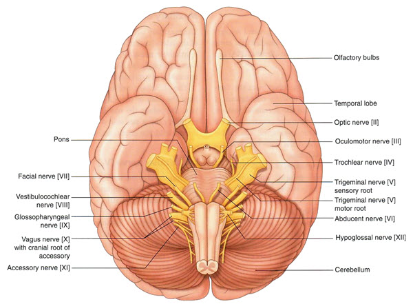
Step 3: Using the Rotate tool, rotate the cadaver to get a sense of how the nerve travels from the lateral surface of the brain stem between the pons and the medula. The nerve has two sets of special afferent fibers. |
 |
Judging by the name of this nerve, what are the functions of these two sets of fibers? |
|
Vestibular = Balance |
|