
IV.B.2 Head: CN V Page 1 of 5
Objectives:3.2.1 Identify the foramen through which the trigeminal nerve exits the cranial cavity and include the components of the trigeminal nerve. 3.2.2 Identify the ophthalmic, maxillary, and mandibular branches of the trigeminal nerve and their branches. 3.2.3 Predict the functional deficits, which would result from damage to cranial nerve V. |
Assigned Readings:Moore's COA: Pages 842-844, 857-861, 911-913, 924, 926, 931, 933-934, 939, 946-947, 953, 961-962, 966, 968-969, 1007, 1011, 1062-1069, 1073-1074. See the blue boxes on pages 1087, 1089 for clinical correlations. See the yellow box on page 1074 for "The Bottom Line." |
Trigeminal CN V
| Step 1: Begin by highlighting the trigeminal nerve. |
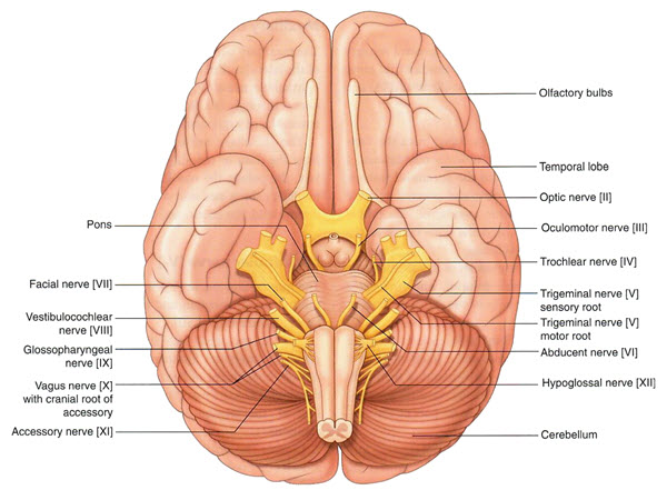
You will notice that the cadaver only demonstrates the portion of the trigeminal nerve as it leaves the pons and moves toward the exit from the cranium.
The trigeminal nerve exits from the anteriolateral surface of the pons as a large sensory root and a smaller motor root. (See drawing above)
As we saw during the demonstration table session from week one, the trigeminal nerve forms a large sensory ganglion prior to exiting the cranium.
 |
The sensory part of the trigeminal nerve divides into three post ganglionic nerves. What are they? |
|
Ophthalmic - V1, Maxillary - V2, and Mandibular - V3 We will discuss each of these in turn. |
|