
IV.B.1 Head: Nasal & Orbital Region Page 1 of 6
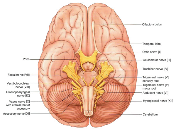
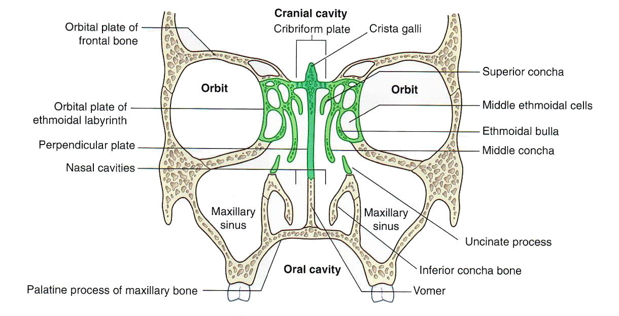
Objectives:3.1.1 Identify the olfactory, optic, oculomotor, trochlear, abducens nerves and their branches. 3.1.2 Identify the foramen through which each of the cranial nerves exit the cranial cavity and include the components of each cranial nerve. 3.1.3 Understand the relationship of the nasal cavity to the anterior fossa, orbits and the cranial nerves. 3.1.4 Predict the functional deficits, which would result from damage to cranial nerves I, II, III, IV, VI. |
Assigned Readings:Moore's COA: Pages 842-844, 858, 898, 904, 908, 969-970, 1062-1068, 1070-1073, 1074-1077. See the blue boxes on pages 1086-1089 for clinical correlations. See the yellow boxes on pages 1070, 1072-1073, 1077 for "The Bottom Line." |
×
![Image]()
Olfactory CN I
| 🔍 Tap on image to enlarge |
 |
 |
 |