
EMT: Anatomy Lab 1: Page 5 of 12
Femoral area
 |
| 🔍 Click on image to enlarge |
×
![Image]()
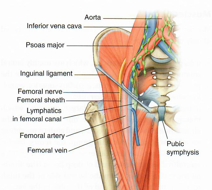
The sequence of structures, moving from lateral to medial forms the NAVEL (Nerve, Artery, Vein, Empty, Lymph) sequence of the structures in the groin. The area below the inguinal ligament where these structures are located is called the femoral triangle.
| Step 27: Add the skin of the groin area to better understand the anatomical landmarks. | |
 |
Step 28: Palpate the femoral artery on your Standardized Patient. |

|
|
 |
Where would you draw blood from the femoral vein? |
|
Medial to where you feel the artery. |
|